English
Sign in
| Price | $7,500.00-$8,300.00 |
| MOQ | 1 |
| Delivery Time | 5 working days |
| Brand | SYSEYE |
| Place of Origin | China |
| Certification | FDA |
| Model Number | RetiCam 3100(Mini) |
| Packaging Details | 380mm(L)x550mm(W)x475mm(H) |
| Payment Terms | T/T |
| Supply Ability | 100 |
| Place of Origin | China | Minimum pupil size | ≥2.8mm |
| Packaging Details | 380mm(L)x550mm(W)x475mm(H) | Dimensions | 380mm(L)x550mm(W)x475mm(H) |
| Power supply | 100-240v 50/60HZ | Field of view | 50° |
| Model Number | RetiCam 3100(Mini) | Supply Ability | 100 |
| Certification | FDA | Weight | 23.5kg |
| Working distance | 17mm | Brand Name | SYSEYE |
| Payment Terms | T/T | feature | Full Automatic |
| Price | $7,500.00-$8,300.00 | Delivery Time | 5 working days |
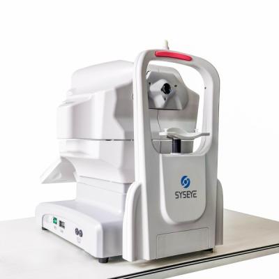
| Product name | Full Automatic Optical Equipments Non Mydriatic Fundus Camera | Minimum Order Quantity | 1 |
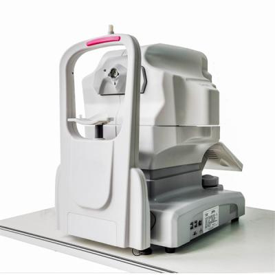
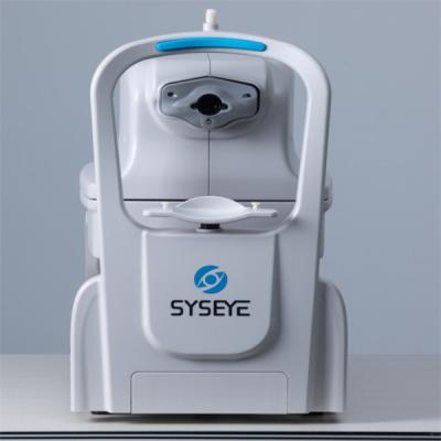
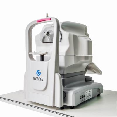
Full Automatic Optical Equipments Non Mydriatic Fundus Camera
RetiCam3100
mini
Non
Mydriatic
Fundus
Camera
can,
by
taking
advantage
of
fundus
imaging
technology
with
light
going
through
pupils,
directly
photography
patients'
retina
and
capture
fundus
images.
It
can
be
used
by
medical
personnel
of
clinical
units,
optometrists
or
health
care
providers
who
master
this
operation.
Applicable
place:
Department
of
Ophthalmology
at
hospitals,
eye
center
and
optometrists'
office.
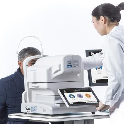
Working principles of Non Mydriatic Fundus Camera
RetiCam 3100, based on near-infrared light imaging technology, can capture the required visible light images of fundus exposed to visible light within milliseconds (no enough time for pupils to shrink).
RetiCam 3100 can photography the retina, i.e. neurosensory tissues of eyes, and then convert the captured optical image into electronic pulses that can be understood by the brain. Pupils serve as both the inlet and outlet of light for illumination and imaging of RetiCam 3100, thus monitoring the fundus. Patients shall put the chin on the chin rest and lean the forehead on the forehead rest. The operator shall operate it manually to focalize and align to the fundus, and capture the fundus images upon clicking, with such images directly shown on the touch screen. Ophthalmologists will trace the progression of eye diseases on the basis of such retinographs, which will serve as the basis of diagnosis and treatment.
|
Acquisition
modes
|
Non Mydriatic Fundus Camera |
|
Field
of
view
|
50°
|
|
Working
distance
|
17mm
|
|
Minimum
pupil
size
|
≥2.8mm
|
|
Focus
modes
|
Auto/manual
|
|
Exposure
modes
|
Auto
/manual
|
|
Operation
|
Auto
/manual
|
|
Photography
|
SLR
camera
|
|
Camera
|
CCD
|
|
Diopter
compensation
|
±25D
|
|
Fixation
|
External
fixation
/
internal
fixation
|
|
Autofocus
assist
light
|
Infrared
LED
|
|
DICOM
3.0
|
Yes
|
|
Customization
AI
port
|
Yes
|


Focusing on the two major public health problems of adolescent myopia and elderly ophthalmology, our company researches new diagnostic and treatment equipment for ophthalmology, develops low-cost applicable technology products, realizes industrialization, reduces the cost of social medical and health system, and serves the strategic needs of national health.The company always adhere to the "high-tech,new vision" for the enterprise development concept.We have a strong technical force, has been awarded 11 patents, and obtained 13,485 quality system certification.Bio has established an efficient marketing team and a perfect after-sales service system to provide medical equipment with the highest cost performance and meticulous service.